MRI-derived risk stratification for early Alzheimer's research.
NeuProScan investigates MRI biomarkers that may indicate elevated risk earlier in the disease trajectory, helping research teams prioritize PET confirmation, enrich cohorts, and track longitudinal change.
For research use only. Not cleared for clinical diagnosis.Why an MRI predictor is useful.
Newly approved anti-amyloid therapies such as Lecanemab and Aducanemab are typically confirmed with amyloid PET imaging, which can be costly and resource-intensive. MRI is widely available and less expensive, yet early-stage detection can be variable. Literature reports false-positive and false-negative rates in the ~27-29% range for early Alzheimer's (Cochrane review).
Research framing
NeuProScan is positioned as a research tool to improve MRI-based risk stratification, reduce unnecessary PET utilization, and identify cohorts for deeper evaluation.
Planning ahead
Earlier signals can support long-term planning for patients and care teams while further diagnostics are scheduled.
Trial readiness
Research cohorts benefit from earlier identification of likely converters for clinical trial recruitment.
Results snapshot
Retrospective evaluation on a cohort labeled healthy at baseline suggests the model can flag a large share of later converters. Performance varies by site and protocol and should be validated per deployment.
- Baseline clinician assessment labeled patients as cognitively healthy.
- Roughly one-third developed Alzheimer's within follow-up windows.
- NeuProScan flagged about 88% of later converters in the retrospective analysis.
These results are research findings and do not represent a cleared diagnostic claim. Prospective validation is required for clinical use.
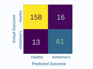
Confusion matrix from retrospective evaluation.
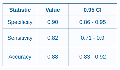
Confidence interval summary of key metrics.
Workflow and platform context
NeuProScan provides a configurable AI workflow for MRI ingestion, feature extraction, and risk scoring. It supports on-device and cloud deployments and allows teams to iterate on custom model variants.
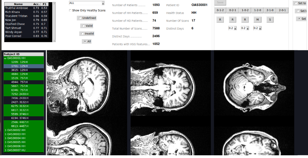
Sample interface showing MRI ingestion and review workflow.
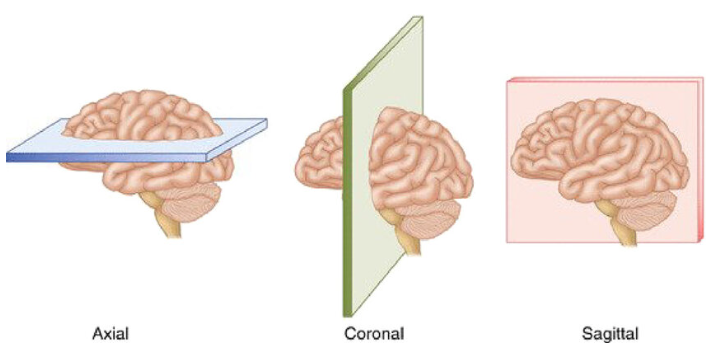
Axial, coronal, and sagittal planes used for model input selection.
Slice selection strategy
Each MRI scan includes hundreds of slices. NeuProScan selects a focused subset across axial, coronal, and sagittal planes to balance signal capture with model efficiency. Current experiments use up to nine slices, including central and adjacent views, to capture structural change while limiting noise.
Discuss model variantsComparative analysis
Benchmarking across internal baselines helps contextualize performance and guide iteration.
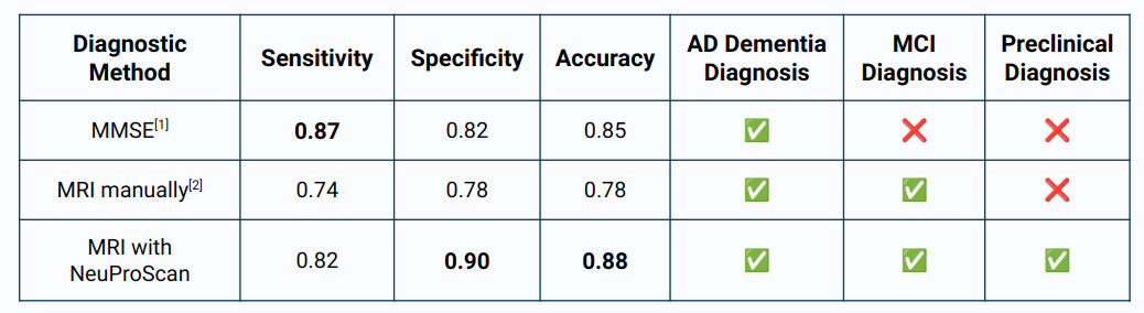
Comparative results across model variants and baselines.
Explore the MRI predictor workflow
Collaborate with HermesLabsIO to evaluate MRI-based risk stratification workflows and tailor models for your research program.